Editor’s Note
This article highlights a promising breakthrough in medical imaging technology. Researchers have developed a new, more accessible method for fabricating microscopic shape-shifting probes that could one day significantly improve MRI scans. By simplifying a previously complex and specialized production process, this innovation may help accelerate the development and future application of these experimental tools.
Microscopic magnetic probes that change shape in response to their environment may greatly enhance magnetic resonance imaging (MRI). However, producing the probes, which are still experimental and have not yet been used in humans, has required access to a cleanroom and expertise in nanofabrication, limiting their widespread use.
Now, researchers at the National Institute of Standards and Technology (NIST) have taken these shape-shifting probes, known as geometrically encoded magnetic sensors, or GEMS, a step further by unveiling a novel fabrication method that is not only faster and cheaper but eliminates the need for specialized instruments.
The scientists reported their work online Dec. 19 in ACS Sensors.
NIST scientists Gary Zabow and Samuel Oberdick, and their colleagues, focused their efforts on building GEMS shaped like tiny hollow cylinders because that shape can be easily fabricated with a mold. For their master mold, the scientists constructed an array of hollow cylinders made of hard silicon, each only about 100 micrometers in diameter — about ten times larger than a red blood cell.
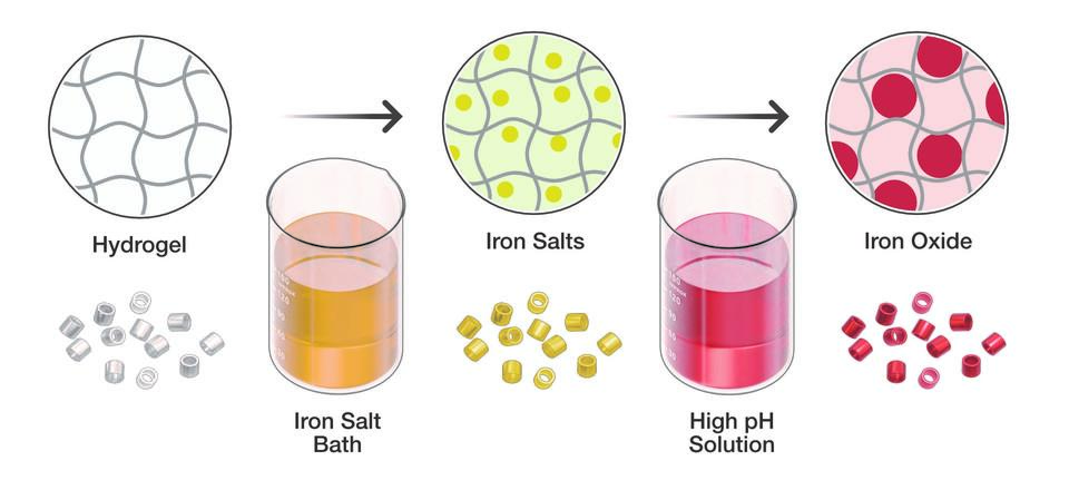
A hard silicon master mold is used to create a flexible polymer mold, which is flipped over and filled with hydrogel. The hydrogel is then cured with UV light, producing the cylindrical microparticles.
The team then demonstrated how researchers with such a master mold could complete the multi-step fabrication process. First, the scientists made a soft-mold “negative” of the master by pouring a liquid polymer on top of the hard silicon mold, allowing it to solidify, and then peeling it off. This created a pliable mold with an array of cylindrical hollow cavities.
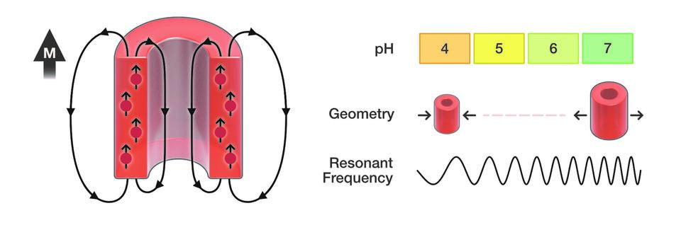
In the next step, the scientists filled each cavity with a liquid precursor to a hydrogel–a network of cross-linked polymers that can absorb large amounts of water. The hydrogel, which had been engineered to shrink or swell in response to changes in acidity or other properties in its microenvironment, is a key component of the GEMS. Engineered hydrogels are inexpensive and easy to make.
After hardening the hydrogels by exposing them to ultraviolet light, the NIST team turned them out of their soft mold, similar to popping ice-cubes out of a silicon tray. The cylindrical hydrogels were then immersed in a bath of iron salts and transferred to a basic solution, which converted the iron salts absorbed by the hydrogels into magnetic oxide particles.
When placed in a strong external magnetic field (labeled M), the iron oxide particles become magnetized, causing the microparticles to develop their own local magnetic field. The microparticles shrink and swell with changes in acidity, which strengthens or weakens this local field and therefore how much the field influences the resonant frequency of protons during an MRI scan.